
OsteoFab® Rx
Everything you need to bring a personalized PEKK implant case from a surgeon conversation to the O.R. — the case checklist, current forms, sales collateral and the evidence behind the technology, in one place.
Questions on a live case? osteofab@oxfordpm.com
Four things a case needs
OsteoFab® Rx devices are custom devices prescribed by a physician for one patient. A complete submission is what keeps the timeline short — here is exactly what to collect.
Prescription
The surgeon confirms no commercially available device is suitable and signs the Rx form.
- Patient identifier, surgery date, side
- Indication and justification for a custom device
- Device description or sketch
Imaging
A thin-slice CT of the affected anatomy is the design input. Contralateral CT and recent X-rays help.
- Scan to the OsteoFab CT parameters
- DICOM export
- Upload, or mail media to OPM
Planning & approval
OPM engineers segment the anatomy, then hold a virtual planning session with the surgeon. Iterations continue until the surgeon approves the design.
- Book the surgeon for a 30-minute call
- Fixation preference and instruments
- Surgeon sign-off on the final design
Manufacture & deliver
The implant is laser-sintered from OXPEKK® powder, inspected and shipped ahead of the surgery date.
- Confirm ship-to and hospital contact
- IFU travels with the device
Typical time from CT receipt to delivery, assuming a complete submission and prompt design approval. Flag urgent limb-salvage or oncology cases to the design team when you submit.
Sell the technology
Current pieces only. Each card shows its release date so you always know you are sharing the latest version.
OsteoFab® Rx brochure
Material technology, the case process and foot & ankle case examples. The primary leave-behind for surgeons.
Foot & ankle case examples
TTC fusion, distal tibial cage, total talus and more — imaging and device photos for each.
Technology slides
Presentation deck covering the OsteoFab® platform, properties and data. Use for lunch-and-learns and VAC meetings.
Custom case examples
Broader application landscape: pelvis, sternum, intercalary segments, cones and wedges.
Surface wicking demo
Thirty seconds that shows what a spec sheet can’t: fluid climbing the printed surface against gravity.
Controlled documents
Document numbers and revisions are shown so a form can be checked against the copy in a surgeon’s office. Superseded revisions should not be used.
| Document | Number | Rev | Use | |
|---|---|---|---|---|
| OsteoFab® Custom Device Prescription Form (US)Physician prescription and justification for a custom device | QMSF-1400 | C | Required · every case | Download |
| OPM Recommended CT Scanning ParametersGive to the imaging center before the scan | QMSD-1400 | B | Required · imaging | Download |
| How to Send Us a CT ScanUpload and mailing instructions for DICOM data | — | 1 | Guide | Download |
| OsteoFab® Custom Device Instructions for UseShips with every device; share with the O.R. team in advance | QMSL-1400 | D | IFU | Download |
| Total Talus — Indications for Custom Device ExemptionLanguage to support the CDE justification for total talus cases | — | — | Reference | Download |
| OsteoFab® Custom Device Inquiry Form (EU + UK)For enquiries outside the US | QMSF-1500 | A | OUS only | Download |
Why PEKK
The thirty-second answer to “why not titanium?” — with the supporting literature under each property, one click from the paper.
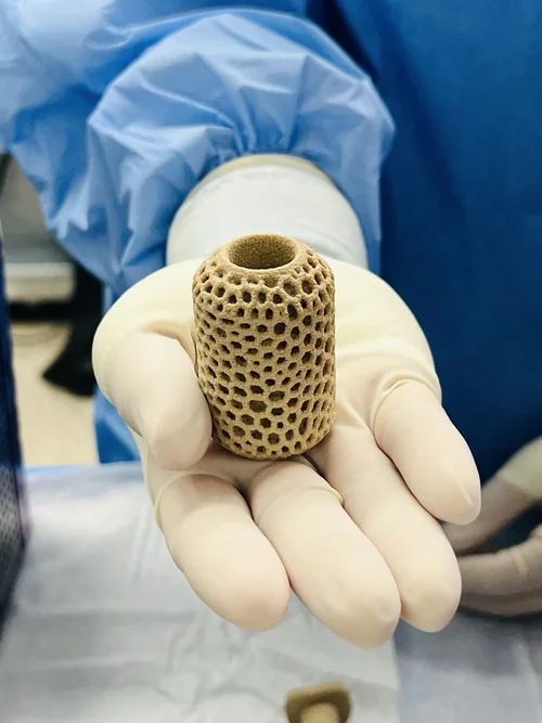
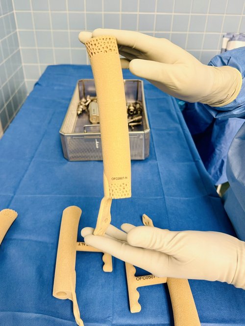
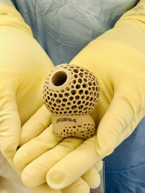
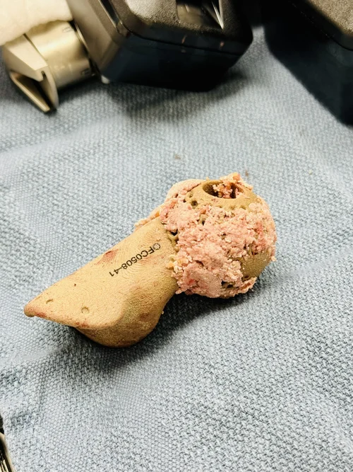
Fluid wicks against gravity across the as-printed surface, seeding the whole implant with the proteins needed for healing.
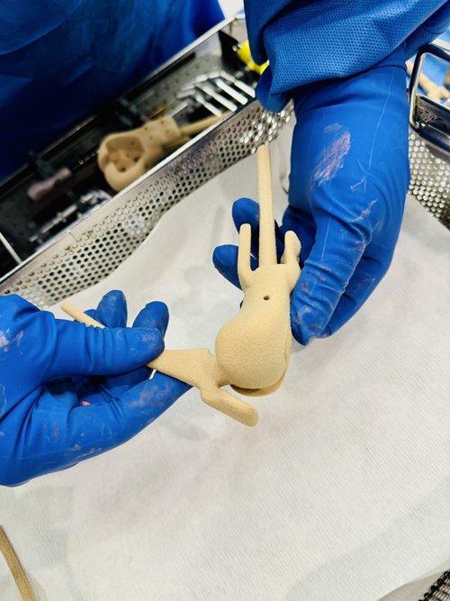
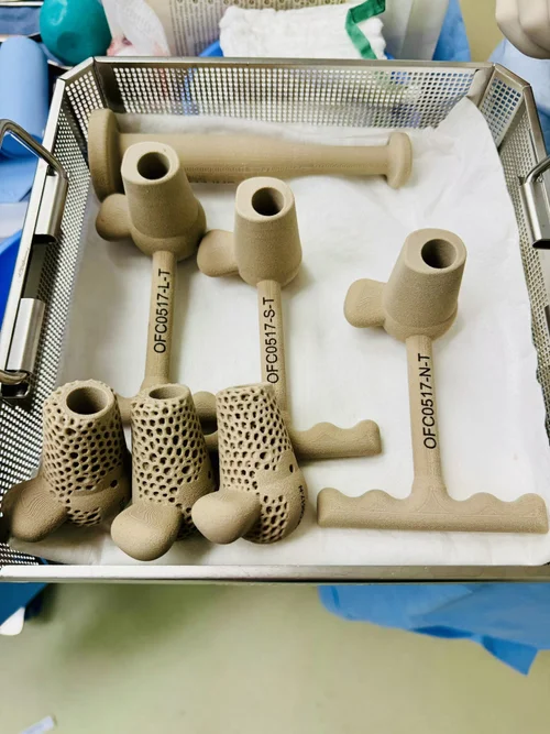
Request a demo sample
Sample requests go to sales@oxfordpm.com. Expect a reply within one business day. Demo implants and material coupons are non-sterile demonstration items and are not for implantation. Please do not include patient names or MRNs — case details belong in osteofabrx.com.
Need pricing?
For a case-specific quotation or a standing price list, email the sales team with the anatomy, indication, proposed surgery date and, if the case is already submitted, the osteofabrx.com case ID.
Email sales@oxfordpm.com